Facing Fungus
What are the diagnostic implications of the alarming rise in fungal infections?
The prevalence of fungal infections is rising worldwide and those more invasive in nature are of great concern (1). Though superficial fungal skin infections, such as ringworm, are easily treatable, invasive deep tissue fungal infections (IFIs) can lead to high morbidity and mortality rates (2).
Two million people are diagnosed with acute IFIs each year and three million people – often those with weakened immune systems – face more severe and chronic fungal infections (3). Some of the most common IFI pathogens include Candida, Cryptococcus, and Aspergillus. Additionally, endemic fungal species like Pneumocystis, Blastomyces, Histoplasma, Paracoccidioides, and Coccidioides have also been implicated in causing severe systemic infections in immunocompromised patients (4,5,6). The US Centers for Disease Control and Prevention (CDC) surveillance data has also demonstrated mortality rates of 25 percent for patients with candidemia and from 40 to 90 percent for those with invasive aspergillosis (IA) – primarily for immunocompromised individuals. All vulnerable and immunocompromised groups, such as organ or hematological transplant recipients, AIDS patients, diabetics, burn victims, and those with chronic respiratory conditions are most at risk of opportunistic IFIs.
The emergence of multi-drug resistant fungal pathogens is certainly cause for concern, with some fungi appearing that are resistant to all types of antifungal medications, either naturally or through exposure (7). In response, clinicians and laboratory staff must remain vigilant, as patient symptoms can often resemble those of non-fungal infections. There should be an increase in diagnostic assessments to promptly and accurately identify the specific fungi causing infection to ensure better patient outcomes and address public health risks (8).
However, diagnosing IFIs is time consuming. Clinical labs often have to test for more frequent viral or bacterial pathogens before they can begin fungal testing. Given the increase of invasive fungal infections, maybe it’s time for labs to adjust testing algorithms for efficient screening and diagnostic protocols.
Fungal public health threats
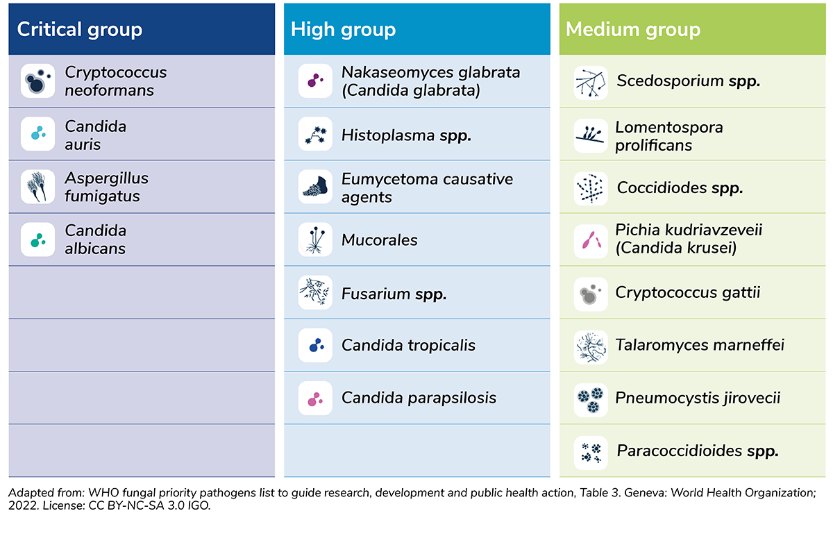
In 2022, WHO released its first list of priority fungal pathogens to highlight the importance in regards to health and creating pathways for patient management (see Figure 1) (9). Four fungal pathogens were rated as critical: Aspergillus fumigatus, Candida albicans, Candida auris, and Cryptococcus neoformans. Seventy percent of IFIs were classified as invasive candidiasis, followed by 20 percent cryptococcosis and 10 percent aspergillosis. This rise in fungal infection is traced to global warming, rises in international travel, and increased drug resistance rates.
The C. auris challenge
First reported in 2009, Candida auris (C. auris) is considered a substantial global threat, frequently multidrug-resistant, with some strains resisting almost all available treatments (10, 11). Notably, 90 percent of C. auris isolates resist fluconazole, 30 percent resist amphotericin B, and less than five percent resist echinocandins. Furthermore, resistance to echinocandins is increasing, with cases tripling year on year in 2021 (12).
C. auris is also associated with high mortality rates – one in three patients die within a month of being diagnosed with the infection (13). It’s often detected in healthcare facilities, persisting on surfaces and transmitting through breathing and feeding tubes. Like other IFIs, immunocompromised patients are the most vulnerable to the dangers of the C. auris infection, but healthy individuals may become colonized without symptoms.
Healthy colonized individuals (asymptomatic carriers) must be identified to evaluate the risk of acute infection and prevent potential spread in healthcare facilities. Precautionary measures are key with patients known to have C. auris, including patient isolation, personal protective equipment, additional sterilization measures, and educating visitors. Advanced information regarding admitted individuals who were previously positive or exposed to C. auris is also pivotal in managing and controlling the spread of disease.
To identify colonized patients, healthcare providers should perform colonization screening – taking a composite swab sample from the patient’s skin near the armpits and groin, and submitting it for lab testing. Clinical testing with blood or urine must be performed if a patient shows symptoms of an infection with an unknown cause.
Whether screening or diagnosing IFIs, a fast-testing turnaround time with accurate identification is essential for positive patient outcomes. Several approaches for C. auris can be seen in Figure 2.

C.auris identification
Culture based phenotypical and biochemical fungal tests take substantial hands-on time of up to 10 days to identify fungal species, and in some cases C. auris may be misidentified as yeast through this testing method (14). However, a recently described novel chromogenic agar (CHROMagar Candida Plus) has promising utility for rapid identification and differentiation of C.auris from other Candida species.
Positive blood culture identification for Candida species is less than 50 percent accurate for bloodstream infections and less than 20 percent accurate for intra-abdominal candidiasis (15). Microscopy and histopathology methods require significant expertise and may not be feasible in all labs. However, while detecting fungal antigens could be affordable, user-friendly, and potentially applicable at the patient’s bedside, there are currently no immunoassays available for C. auris. Additionally, limited sensitivity has restricted development of commercially available immunochromatographic assays for yeast detection (16).
Mass spectrometry techniques, such as matrix-assisted laser desorption/ionization-time of flight (MALDI-TOF), can differentiate C. auris from other Candida species, but not all reference databases with these devices allow C. auris identification. There are FDA-cleared commercial MALDI-TOF systems with updated libraries that allow for C. auris identification with quick results, but the initial cost of equipment and training personnel are high.
Molecular tests
Molecular tests provide the promise of accurate alternatives for fungal pathogen detection. Several syndromic or PCR pathogen panel tests are FDA approved and include more than 40 targets associated with bloodstream infections – such as gram-negative bacteria, gram-positive bacteria, yeast with C. auris, and antimicrobial resistance genes – allowing for pathogen identification as quickly as an hour from positive blood culture. Other major advantages of these tests include sample-to-answer automation and a high number of targets evaluated in a single test.
Targeted laboratory PCR methods have been reported for detecting C. auris, but there are no commercially available FDA-cleared PCR tests for identifying colonized, asymptomatic patients (17). However, PCR has demonstrated improved sensitivity in environmental screening for isolates across, for example, New York healthcare facilities (18).
Importantly, colonization cannot be cured using antifungal medications, and treatment is not required or recommended for people found to be carrying C. auris without any symptoms or signs of infection. Commercial FDA-cleared PCR tests highly specific and sensitive to C. auris detection will therefore be key to preventing the spread from colonized individuals.
Overall, rapid molecular testing offers an accurate alternative to traditional fungal pathogen testing. PCR-based methods effectively detect fungal strains, even when culture and other tests fail – allowing for timely clinical action. Multiplex panels in PCR testing enable labs to identify various pathogens while distinguishing between similar strains often missed in standard fungal tests. Additionally, molecular tests can pinpoint genetic markers of drug resistance for aiding treatment decisions.
When selecting or developing a PCR method to validate, it’s important to consider multiple factors: sensitivity and specificity, lab capacity and equipment, clinical evaluation with appropriate sample type, cost, and turnaround time for swiftly identifying colonized patients.
Looking forward, the rising incidence of emerging fungal infections call for clinical labs to plan for increased fungal testing for screening and diagnosis. Molecular tests can provide labs with accurate scalability, cost-effectiveness, and less hands-on time than traditional approaches for approaching these tasks. Specifically in the US, FDA cleared C. auris PCR tests will be pivotal for broader lab availability to identify colonized patients on a global scale and respond to this growing health threat.
References:
Centers for Disease Control and Prevention, “Fungal disease awareness week” (2023). Available at: www.cdc.gov/fungal/awareness-week.html.
L Pagano et al., “Invasive fungal infections in high-risk patients: report from TIMM-8 2017,” Future Sci OA, 4, (2018). PMID: 30057784.
F Bongomin et al., “Global and multinational prevalence of fungal diseases-estimate precision.” J Fungi (Basel), 3, (2017). PMID: 29371573.
TR Malcolm et al., “Endemic mycoses in immunocompromised hosts.” Curr Infect Dis Rep, 15, (2013). PMID: 24197921.
J Delaloye et al., “Invasive candidiasis as a cause of sepsis in the critically ill patient.” Virulence, 5, (2014). PMID: 24157707.
S Tsay et al., “Burden of Candidemia in the United States, 2017.” Clin Infect Dis, 71, (2020). PMID: 32107534.
J Houšť et al., “Antifungal Drugs.” Metabolites,10 (2020). doi.org/10.3390/metabo10030106.
W Fang et al., “Diagnosis of invasive fungal infections: challenges and recent developments.” J Biomed Sci, 30 (2023). PMID: 37337179.
World Health Organization, “WHO releases first-ever list of health-threatening fungi” (2022). Available at www.who.int/news/item/25-10-2022-who-releases-first-ever-list-of-health-threatening-fungi.
K Satoh, et al., “Candida auris sp. nov., a novel ascomycetous yeast isolated from the external ear canal of an inpatient in a Japanese hospital.” Microbiol Immunol, 53, (2009). PMID: 19161556.
S Vallabhaneni, et al., “Investigation of the First Seven Reported Cases of Candida auris, a Globally Emerging Invasive, Multidrug-Resistant Fungus-United States, May 2013-August 2016.” Am J Transplant, 17, (2017). PMID: 28029734.
Centers for Disease Control and Prevention, “Increasing Threat of Spread of Antimicrobial-resistant Fungus in Healthcare Facilities” (2023). Available at: www.cdc.gov/media/releases/2023/p0320-cauris.html.
Centers for Disease Control and Prevention, “Fact sheets” (2023). Available at: www.cdc.gov/fungal/candida-auris/fact-sheets/index.html.
C Keighley et al., “Candida auris: Diagnostic Challenges and Emerging Opportunities for the Clinical Microbiology Laboratory.” Curr Fungal Infect Rep, 15, (2021). PMID: 34178208.
CJ Clancy et al., “Non-Culture Diagnostics for Invasive Candidiasis: Promise and Unintended Consequences.” J Fungi (Basel), 4, (2018). PMID: 29463043.
S Otašević et al., “Non-culture based assays for the detection of fungal pathogens.” J Mycol Med, 28, (2018). PMID: 29605542.
EK Dennis et al., “So Many Diagnostic Tests, So Little Time: Review and Preview of Candida auris Testing in Clinical and Public Health Laboratories.” Front Microbiol, 12, (2021). PMID: 34691009.
E Adams et al., “Candida auris in Healthcare Facilities, New York, USA, 2013–2017.” Emerg Infect Dis, 24, (2018). PMID: 30226155.
